Designed by Dr. Piercarlo Meinero, VAAFT is indicated in the surgical treatment of complex anal fistulas and their recurrences.
The technique is performed under direct vision, thanks to the use of a fiber optic device, the Meinero's Fistuloscope (manufactured by Karl Storz SE & Co - Tuttlingen - Germany) which is connected to a video system and a bag of irrigation solution. A monopolar electrode, connected to a power unit, an endo-brush and a thin endo-forceps complete the surgical kit.

VAAFT (Video Assisted Anal Fistula Treatment)

These "accessories" pass through the fistuloscope which has an optical channel for the vision, and an operating channel for the passage
both of the instruments, and the washing solution. The internal orifice of the fistula can be closed in various ways: using a mechanical stapler, a particular suture (flap) or using a small piece of the patient's dermis (Autologous Dermis Flap).
both of the instruments, and the washing solution. The internal orifice of the fistula can be closed in various ways: using a mechanical stapler, a particular suture (flap) or using a small piece of the patient's dermis (Autologous Dermis Flap).

VAAFT Description

The VAAFT includes a diagnostic phase and an operational phase.
A) The Diagnostic Phase
Aim of this phase are the correct definition of the fistula "anatomy". It is mandatory to recognize not only the correct direction of the fistula pathway, but also any secondary tracts or possible abscess cavities. It is also essential to identify the correct location of the internal fistula opening.
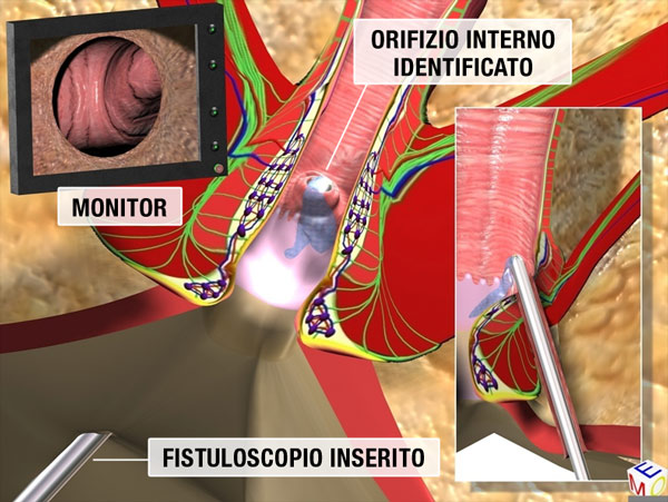
The fistuloscope is inserted through the external orifice and, thanks to the continuous flow of the 1% glycine and mannitol solution, the fistula pathway clearly appears on the screen. Using slow movements, the Surgeon reaches the end of the fistula main tract, which is the internal fistula opening, in the most of cased located into the anal canal or into the rectum (figure 2).
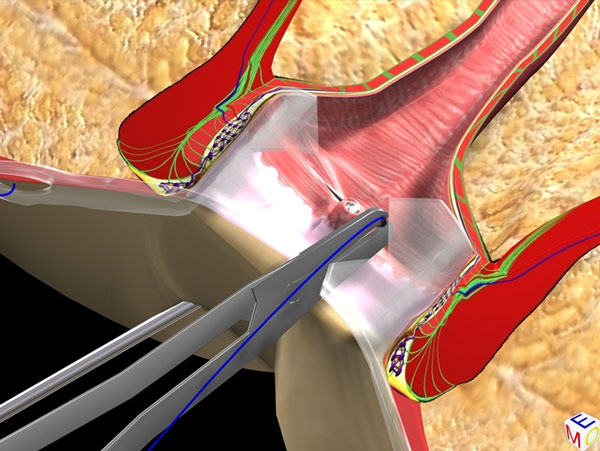
The progression of the fistuloscope through the fistula pathway, can be likened to that of a car in a tunnel. If other secondary tracts or abscess cavities are visible, the Surgeon can easily highlight and treat them, always visually. Once identified, the internal orifice is isolated by applying two or three stitches on it, but without knotting them, just to don't lose it during the next phase (figure 3)
Aim of this phase are the correct definition of the fistula "anatomy". It is mandatory to recognize not only the correct direction of the fistula pathway, but also any secondary tracts or possible abscess cavities. It is also essential to identify the correct location of the internal fistula opening.
The fistuloscope is inserted through the external orifice and, thanks to the continuous flow of the 1% glycine and mannitol solution, the fistula pathway clearly appears on the screen. Using slow movements, the Surgeon reaches the end of the fistula main tract, which is the internal fistula opening, in the most of cased located into the anal canal or into the rectum (figure 2).
The progression of the fistuloscope through the fistula pathway, can be likened to that of a car in a tunnel. If other secondary tracts or abscess cavities are visible, the Surgeon can easily highlight and treat them, always visually. Once identified, the internal orifice is isolated by applying two or three stitches on it, but without knotting them, just to don't lose it during the next phase (figure 3)
B) The Operative Phase
Aims of this phase are the fistula "eradication" from within, the removal of the necrotic tissue, and the hermetic closure of the internal fistula opening.
Aims of this phase are the fistula "eradication" from within, the removal of the necrotic tissue, and the hermetic closure of the internal fistula opening.
The monopolar electrode, connected to the high-frequency electrosurgical generator, is introduced through the operative channel. The Surgeon can clearly see on the screen the spherical tip of the electrode, and can start cauterizing the internal wall of the fistula, always under vision.
The continuous infusion of 1% glycine and mannitol solution allows not only an excellent vision, but also the flowing out of the necrotic material, produced during the fistula cauterization, into the rectum through the internal orifice which was previously left open.
The surgeon "destroys" the entire fistulous tract, but also any possible secondary tract and/or abscess cavities (figure 4). Using an endo-brush introduced through the operating channel, the perfect cleaning of the fistula is done, always visually.
The continuous infusion of 1% glycine and mannitol solution allows not only an excellent vision, but also the flowing out of the necrotic material, produced during the fistula cauterization, into the rectum through the internal orifice which was previously left open.
The surgeon "destroys" the entire fistulous tract, but also any possible secondary tract and/or abscess cavities (figure 4). Using an endo-brush introduced through the operating channel, the perfect cleaning of the fistula is done, always visually.

The internal opening closure
A) With a Stapler
At this point the fistuloscope is removed, and the internal fistula opening must be closed. The Surgeon needs to choose from various options the one that is suited best to close the internal opening. In patients without a prior history of anorectal surgery, the surrounding tissue is normally relatively soft and free of scars. So, the mucosa can be easily raised by applying traction on the two stitches placed during the diagnostic phase, by obtaining a sort of "volcano-shaped" eminence. In these cases, a mechanical stapler (linear or semi-circular) can be applied to the base of this eminence to achieve a hermetical closure of the internal opening (Fig. 5 and 6).
A) With a Stapler
At this point the fistuloscope is removed, and the internal fistula opening must be closed. The Surgeon needs to choose from various options the one that is suited best to close the internal opening. In patients without a prior history of anorectal surgery, the surrounding tissue is normally relatively soft and free of scars. So, the mucosa can be easily raised by applying traction on the two stitches placed during the diagnostic phase, by obtaining a sort of "volcano-shaped" eminence. In these cases, a mechanical stapler (linear or semi-circular) can be applied to the base of this eminence to achieve a hermetical closure of the internal opening (Fig. 5 and 6).


However, when the patient underwent surgery several times (multi-recurrence), the margins of the internal orifice are too tough and
sclerotic. So the use of a mechanical stapler can be difficult. In those cases, we use other strategies.
B) Advancement FlapI
Closure of the internal orifice can thus be obtained by means of a hand-made suture after removal of the scar tissue. A section of fullhealthy mucosa surrounding the internal orifice is mobilized and used to "cover" the internal orifice (advancement flap).
C) ADF: Autologous Dermis Flap
Approximately three years ago, we have been proposing an additional method to close the internal opening. An adequate size of the patient's dermis is harvested from the outermost margin of the external opening. The mucosa is incised at the internal opening and elevated to expose the underlying inner muscular layer. A single stitch is made to pass through the dermis graft which is then secured over the inner muscular layer, precisely on the internal opening. This autologous dermis graft, which acts both as a barrier and scaffold for tissue repair, provides for an airtight closure of the internal opening. The mucosa is finally closed with separate stitches.
sclerotic. So the use of a mechanical stapler can be difficult. In those cases, we use other strategies.
B) Advancement FlapI
Closure of the internal orifice can thus be obtained by means of a hand-made suture after removal of the scar tissue. A section of fullhealthy mucosa surrounding the internal orifice is mobilized and used to "cover" the internal orifice (advancement flap).
C) ADF: Autologous Dermis Flap
Approximately three years ago, we have been proposing an additional method to close the internal opening. An adequate size of the patient's dermis is harvested from the outermost margin of the external opening. The mucosa is incised at the internal opening and elevated to expose the underlying inner muscular layer. A single stitch is made to pass through the dermis graft which is then secured over the inner muscular layer, precisely on the internal opening. This autologous dermis graft, which acts both as a barrier and scaffold for tissue repair, provides for an airtight closure of the internal opening. The mucosa is finally closed with separate stitches.
THE COMPANIES WE WORK WITH

